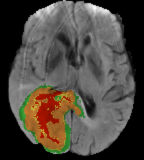
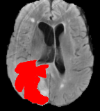
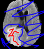
Manual

The completely manual method for segmentation would be to draw the desired borders directly onto the raw image. This would take too much time and be prone to errors, especially due to fatigue. Manual segmentation requires the radiologist to use the visual and spatial information presented by the MRI images along with anatomical and physiological knowledge gained through training and experience. Procedure involves the radiologist going through multiple slices of images slice by slice, diagnosing the tumor and manually drawing the tumor regions carefully.

Issues with Manual Annotation

Apart from being a time consuming task, manual segmentation is also radiologist dependent and segmentation results are subject to large intra (variability between repeated administrations of segmentation by a single rater) and inter rater variability (variability between repeated administrations of segmentation by different raters)[3]

Intraoperator Repeat Results:

  • 95%
  • 94%
  • 85%

Interoperator comparisons:

  • 94%
  • 93%
  • 81%

Given the vital nature of accuracy in this scenario it's a suprising that the variation is so high, and suggests that automated solutions will probably be far more consistent.

Using computers to help plan radiotherapy could help deliver better treatment for patients by speeding up the process and improving accuracy.
Dr Justine Alford, senior science information officer at Cancer Research UK

Annotation

General segmentation of the human brain involves defining structures by borders correspodning to signal intensity transitions at Brain-Cerebral Signal Fluid or gray-white matter interfaces.

  • Necrosis (Red): Dead cells from the tumor
  • Edema (Green): Brain swelling due to new blood vessels growing in and near the tumor
  • Non-enhancing Tumor (Yellow): Tumor cells that aren't inflammatory.
  • Enhancing Tumor (Orange): Uptake of contrast agent - hyper intense on MRI T1Gd - this means that there's an inflammatory process.

Semi-Automatic

Semi-Automatic methods require interaction of the user for three main purposes:

  • Initialisation - Defining a region of interest (ROI) containing the approximate tumor region before the automatic algorithm acts and the parameters of pre-processing methods can also be adjusted to suit the input images
  • Intervention - Automated algorithms can be steered towards a desired result during the process by receiving feedbacks and providing adjustments in response.
  • Feedback response and evaluation - Evaluate the results and modify or repeat the process if not satisfied.

Thresholding

The simplest method of image segmentation that directly dicides the image gray scale information processing based on the gray value of different targets. Threshold segmentation can be divided into a local threshold method and global threshold method. The most commond threshold algorithm is the "largest interclass variance method" which selects a globally optimal threshold by maximising the variance between classes.

The global threshold method divides the image into two regions of the targt and the background by a single threshold.[5]

The local threshold method needs to select multiple segmentation thresholds and divide the image into multiple regions and backgrounds.

Advantages

  • Calculation is simple
  • Operation speed is fast
  • In particular, when the target and the background have high contrast, the segmentation effect is good

Disadvantages

  • Difficult to obtain accurate results where there is no signifant grayscale areas
  • Only takes into account the gray information of the image without considering the spatial information
  • Sensitive to noise and grayscale unevenness

Graph Cut [4]

The image is divided into "object" and "background" segments using a graph cut approach. A graph is formed by connecting all pairs of neighbouring image pixels (voxels) by weighted edges. Certain voxels have to be identifed providing necessary clues about image content. Objective is the cheapest way to cut the edges in the graph so that the object seeds are completely separated from the background seeds. This is a known issue

Place a mark on the image to indicate regions desired as foreground and regions as background. First marks are done on forground and then again on the background. This gives a reasonable first pass on the segmentation but if the intensity values are too similar then a good segmentation can't be achieved

Automatic

Automatic segmentation of gliomas is challenging as tumor bearing MRI data is 3D data where tumor shape, size and location that can vary from patient to patient. Tumor boundaires are often unclear and irregular with discontnuities posing very hard challenges for edge-based methods.Automatic tumor segmentation has the potential to decrease time between diagnostic tests and treatment by providing an efficient and standardized report of tumor location in a fraction of the time it would take a radiologist to do so.

Automatic Methods

Footnotes

  • [1]Havaei, M. et. al, Brain Tumor Segmentation with Deep Neural Networks. arXiv preprint arXiv:1505.03540, 2015.
  • [2]Stupp et al., Effects of radiotherapy with concomitant and adjuvant temozolomide versus radiotherapy alone on survival in glioblastoma in a randomised phase III study: 5-year analysis of the EORTC-NCIC trial. The Lancet Onc., 2009.
  • [3]White D, Houston A, Sampson W, Wilkins G. Intra and interoperator variations in region-of-interest drawing and their effect on the measurement of glomerular filtration rates 1999; 24:177–81.
  • [4]Boykov Y., Jolly MP. (2000) Interactive Organ Segmentation Using Graph Cuts. In: Delp S.L., DiGoia A.M., Jaramaz B. (eds) Medical Image Computing and Computer-Assisted Intervention – MICCAI 2000. MICCAI 2000. Lecture Notes in Computer Science, vol 1935. Springer, Berlin, Heidelberg
  • [5]Davis L S, Rosenfeld A, Weszka J S. Region extraction by averaging and thresholding[J]. IEEE Transactions on Systems, Man, and Cybernetics, 1975 (3): 383-388.
  • [6]Ivana Despotović, Bart Goossens, and Wilfried Philips, “MRI Segmentation of the Human Brain: Challenges, Methods, and Applications,” Computational and Mathematical Methods in Medicine, vol. 2015, Article ID 450341, 23 pages, 2015. doi:10.1155/2015/450341