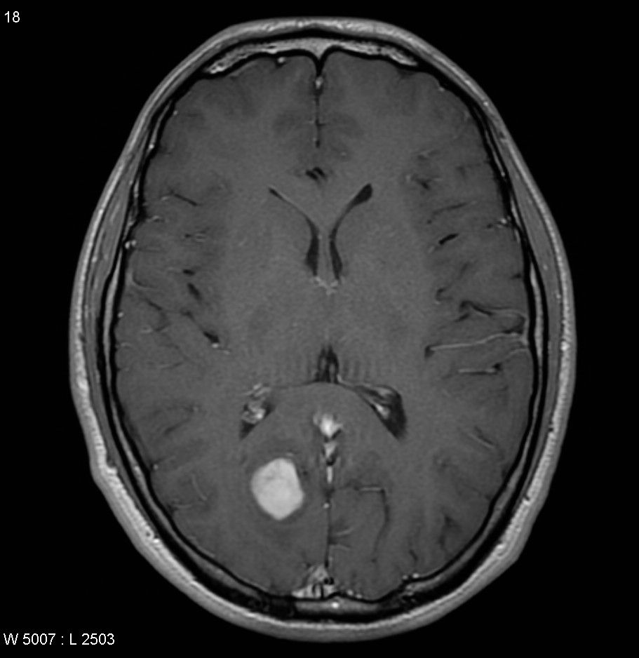
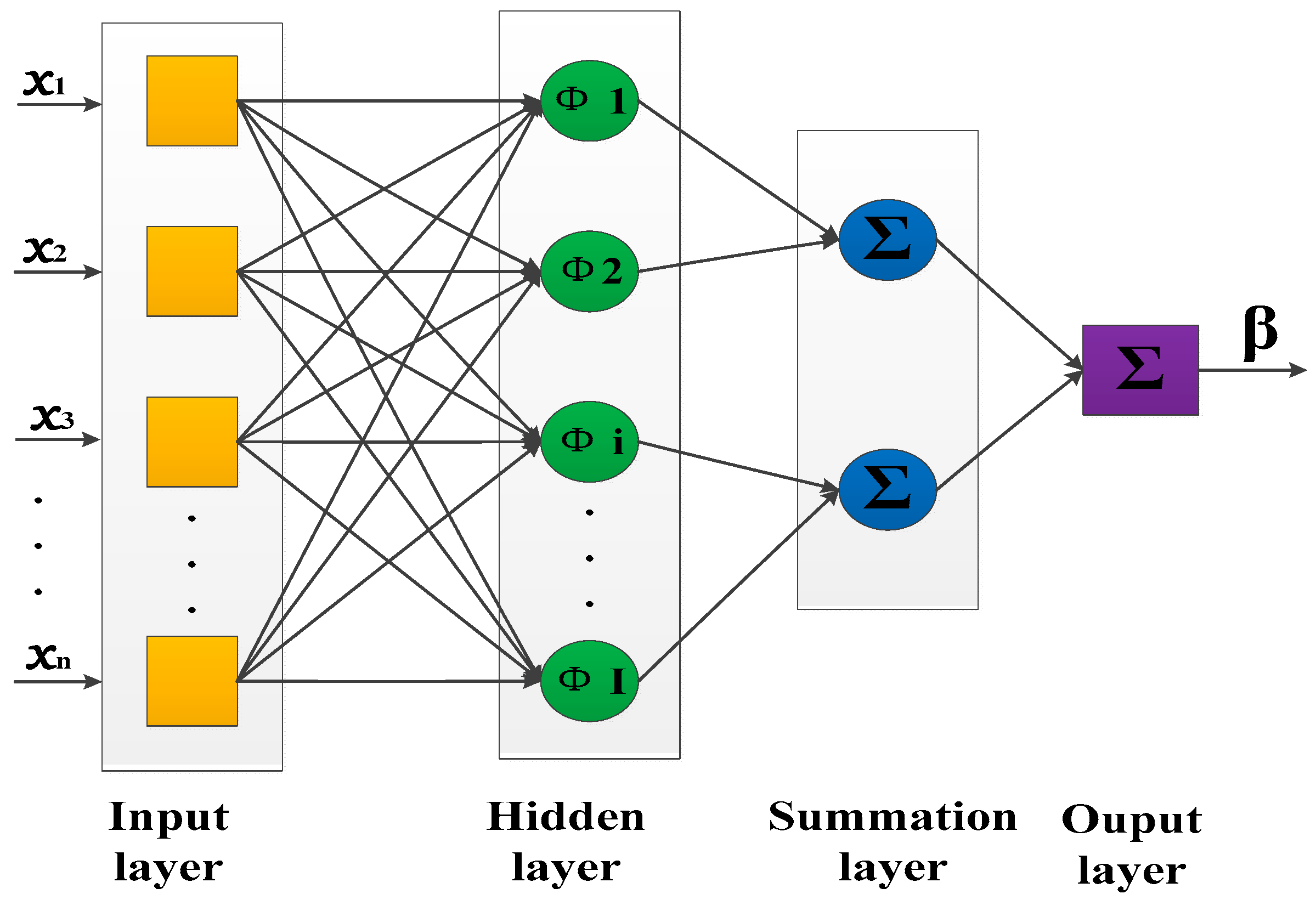
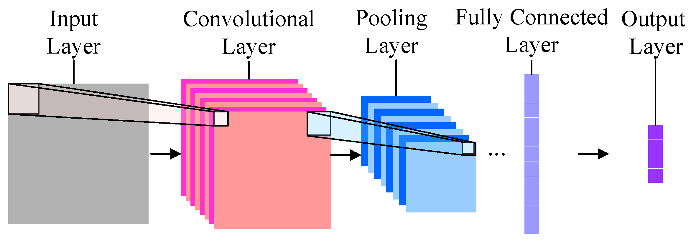
Overview of Brain Tumors and MRIs

Everything a budding medical image analyst would want to know about brain tumours and MRI scans.

Image Pre-processing

The MRI images need manipulating before neural nets or other operations are used in order to ensure they are as clear and compatible as possible. Click below to read about how this is achieved.

Image source: R.Lavanyadevi, M.Machakowsalya, J.Nivethitha, A.Niranjil Kumar, Brain Tumor Classification and Segmentation in MRI Images using PNN, 2017

Image Segmentation

Image segmentation is the process of dividing an image into a set of semantically meaniningful regions. Although done manually and semi-automatically now, there is both need and potential for automatic methods.

Support Vector Machines

SVMs are a type of supervised machine learning method, useful in classification problems. Therefore they have been proposed extensively for classifying brain tumours as malignant or benign from MRI images, showing great accuracy.

Probabilistic Neural Networks

PNNs are becoming very popular with regards to automated classification of brain tumours from MRI scans. They are different to CNNS in that they use radial basis functions and Bayesian probability to classify the input.

CNNs

Convolutional Neural Networks(CNNs) have been developed more recently than PNNs and use a slightly alternate method to classify brain tumours.

About the Creators

Click on the images below to find out more about the people who made this site: